


Northern Blotting is a laboratory technique used to detect and quantify specific RNA molecules within a biological sample. While a Western blot looks at proteins and a Southern blot looks at DNA, the Northern blot tells scientists which genes are actually “turned on” (expressed) in a cell at a specific time. By measuring the amount of messenger RNA (mRNA), researchers can determine if a disease, a drug, or an environmental change is causing a cell to ramp up or shut down the production of specific instructions.
How Northern Blotting Works
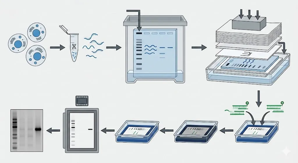
RNA is notoriously fragile and prone to being degraded by enzymes called RNases that are everywhere—even on your fingertips. Because of this, the process requires extreme care and “clean” conditions.
- RNA Separation
Total RNA is extracted from cells and separated by size using gel electrophoresis. Because RNA is single-stranded, it tends to fold into complex shapes; to prevent this, a “denaturing” agent (like formaldehyde) is added to the gel to keep the RNA molecules linear so they move strictly according to their length.
- Transfer
Just like in a Western blot, the separated RNA strands are moved from the fragile gel onto a sturdy nylon or nitrocellulose membrane. This is often done via capillary action, where a stack of paper towels “wicks” SSC (saline-sodium citrate) buffer through the gel and membrane, pulling the RNA along with it and trapping it on the surface of the membrane.
- Hybridization
The membrane is exposed to a probe—a short, single-stranded piece of DNA or RNA that is complementary to the target sequence. This probe is “labeled” with a radioactive atom or a fluorescent dye. The probe scans the membrane and binds (hybridizes) only to its matching RNA partner, ignoring all the other RNA “noise.”
- Detection
After washing away the unbound probes, the membrane is placed against X-ray film or scanned with a digital imager. The labeled probes create dark bands or glowing lines. The thickness and intensity of the band tell the scientist exactly how much of that specific mRNA was present in the original sample.
